


This Is Your Respiratory System
The 1,500 mile journey of every breath you'll ever have.

Welcome to The Feel Good Life! A newsletter about health, prevention, empathy, and hope. Join me, Dr. Mariana, as I explore all sides of good health and a good life. New around here? Get started.

Hi!
In our series 'Basics Matter’ we have covered so far the majestic Nervous System and the hardworking Cardiovascular System—the first one being the control tower, and the second one being the master life provider for the entire human body.
In our next chapter we are covering the system working hand in hand with the heart; the sacred oxygen provider of our entire cellular universe, allowing every single bodily function to happen.
Welcome to the Respiratory System!
In today’s email we are going to focus on:
What is the respiratory system?
The fantastic voyage of a single breath
What does the respiratory system looks like?
How do lungs and heart connect?
Since the beginning of this series, we’ve been learning four main things about each system so that you can understand how your human body works: anatomy, physiology, pathophysiology and prevention—in other words: how it looks like, how it works, what happens when it gets sick, and how to prevent. This is key in preventive medicine. The understanding that a patient can have about their own body increases all chances of them sticking to treatments, guidelines, and lifestyle changes to improve not just the status of a diagnose but overall health in the long run.
And that’s what this series is for.
As we dive into the respiratory system, I will explain things as usual, but instead of really long-form posts, I will intend to make shorter and more digestible bites. If you are new here, let me point you to the previous episodes so you can catch up in your own time:
Let’s begin.

What Is The Respiratory System?
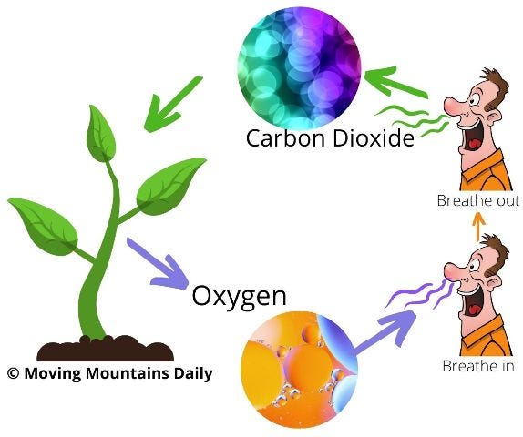
The respiratory system is the body's system in charge of breathing, taking in oxygen as we inhale and releasing carbon dioxide with every exhale. This exchange of oxygen and carbon dioxide is called respiration.
Oxygen is vital for every cells' functioning. And since we’re made up of millions of cells, it becomes then mandatory to have a healthy respiratory system for proper oxygen supply and waste removal.
This system includes the nose, mouth, throat (pharynx), larynx, windpipe (also known as trachea), lungs, diaphragm and other muscles, all working together to move air in and out of the body. This system is essential for 1) providing the body's cells with the oxygen they need to function, and 2) removing the waste gas carbon dioxide—which is used by plants and trees to produce oxygen back into the air we breathe.

The Fantastic Voyage Of A Single Breath
Let’s imagine you are a gulp of fresh air about to enter a person’s body:
As air, you’re invisible, incredibly light, and made gas: oxygen (21%), nitrogen (78%) and traces of carbon dioxide, argon, hydrogen and other substances.
You’re floating past a person just as they take a deep sniff of the air. As they breathe in, the suction from their nose draws you into their body, and your wild journey begins.
Inside the nostrils, you can see and feel tiny hairs brushing against you and through you. These hairs are called cilia and they’re lined along the nose’s inside walls to clean you of any dust particles might go inside and damage a person's lungs. They also warm you up, so your temperature doesn’t feel like a shock to the person's system as you head down.
You’re now travelling towards the back of the nose and into the throat. In here, you can feel it’s warm and humid. You pass a wide opening - that’s the mouth, through which air and food can enter, but not right now. You hurtle into growing darkness, past an odd, pendulous bell-like protrusion hanging from the back of the mouth. That’s the uvula! It's part of the mechanism that stops food getting into people's noses when they eat. Clever stuff! Let’s keep going.
Heading down the long tunnel of the throat, you reach a place where there’s an intersection. On the left, there’s a soft, pink, wet tube where food goes to the stomach. On the right you have a white fence that restricts the movement of everything except air. This permeable fence is called the epiglotis. When an inhalation is happening, the white fence uses air sensors to recognise the presence of nothing but the breath. It opens in front of you, allowing you passage through it.
As you cross through the epiglotis fence, you reach a small section looking like a bridge, called the larynx, better known as the "voice box" for fairly obvious reasons. Beyond here you dive down a tunnel made of many, many rings of cartilage, like you're racing through a multi-segmented subway. You’re now into the trachea - a warm, damp, lane into the lungs, currently operating in one direction only. As you go in, you'll feel no air is coming out, otherwise there would be pressure build-ups and potential accidents aplenty with this sensitive, easily-damaged part of your airway. Instead, it's a peaceful and easy transit.
At the end of the trachea, you see a new intersection: one road to the left and one road to the right. These are the only two entrances, one to each lung. You choose one of them and plunge deeper.
Now you're entering the official landscape of the lungs, an endlessly branching series of tunnels, each sprouting multiple smaller, narrower junctions, like you're inside the trunk of a tree and heading up into the branches. The tunnels’ walls are soft, warm and wet, and sometimes covered in a layer of protective mucus, helping keep them clean of impurities as the air (that's you!) flows inwards.
By the way "you" has now ceased to exist as a single entity. You’re now all over the lungs, as many flowing packets of air absorbed into the vast lung tunnel system. (I really do mean vast. If all these airways were laid out end to end, they'd stretch for 2,400 kilometres (1,500 miles!)
At the farthest end of each tunnel, you reach the ultimate destination of the breath - the alveoli, tiny balloon-shaped bags that process the gases within air. Because of the incredible number of branches of the lungs, there's an equally colossal number of alveoli, anything between 300 to 500 million of them! It's here that the lung tissue touches the circulatory tissue, allowing the oxygen/carbon dioxide exchange to happen. This is how oxygen from the air is taken up by the arteries and transferred into blood cells to race away to feed every cell in the human body.
After this exchange has happened, you have now delivered the oxygen and are now loaded up with carbon dioxide, ready to travel out of the lungs again in the form of an exhale. The opposite journey begins: from the alveoli all the way up, towards the broader branches of the lungs, up into the trachea, through the white fence of the epiglotis, over the larynx bridge, up the throat, passing by the mouth opening, continuing upwards into the back of the nose, into the nostrils and finally - *whoosh* - you've rejoined the outer world as a puff of warm breath, ready to be photosynthesized by plants and trees back into oxygen that's fit for another person's lungs again!
What a journey!
You have now accomplished to deliver one load of vital oxygen into the body and removed one load of carbon dioxide from the lungs into the outside world. You just gave your body one entire breathe of health into every cell. Good job, you.
Here’s a simple visualisation of this breathing journey:

A Closer Look: Upper & Lower Respiratory System
Now, as we mentioned, the respiratory system includes the nose, mouth, throat (pharynx), larynx, windpipe (trachea), and lungs. It also includes several muscles, like intercostal muscles, abdominal muscles, and the diaphragm, to help the lungs expand as we breathe in.
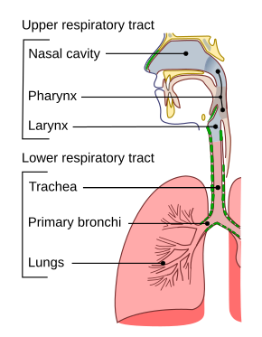
As a quick recap, the air enters the respiratory system through the nostrils (also called nares). In here, air is warmed up and humidified, while tiny nasal hairs called cilia help filter dust particles from entering the lungs. It then travels down through the throat (pharynx), the larynx (voice box), the trachea and finally entering the lungs and its complex tunnel pathways.
The area between the nose/mouth and the larynx, is considered the upper respiratory system. From the trachea all the way down into the alveoli, we have the lower respiratory system. We use this to define the location of respiratory infections.
Every person has two lungs sitting inside their chest, each on one side of the heart, all protected by your rib cage. The lungs are the main structure in the respiratory system.
Doctors who specialise, treat and study the lungs and the respiratory system are called Pneumologists.
The lungs are divided into sections called lobes—two in the left lung and three in the right lung. The first branches as you enter the lungs are called bronchi. As you go in, they narrow up and become bronchioles, and finally, alveoli.
In the alveoli sacks is where the oxygen/carbon dioxide exchange happens.

Other structures important to the respiratory system.
Another set of vital structures that don’t belong inherently to the respiratory system, but impacts it directly, are the sinuses: air-filled spaces in the skull and face that help warm, humidify, and filter the air we breathe. Additionally, they produce mucus, which helps trap dust, germs, and other irritants, protecting the lungs from potential inflammation.
The main sinuses are located exactly on top an on the sides of the nasal cavity and they drain out through the nasal passage. They can become clogged and infected, provoking a well-known condition called sinusitis - (we’ll talk more about it in email 2 as we cover diseases of the respiratory system).
You can see the sinuses here on a side view (image 1) and frontal view (image 2) of the face:


Muscles also play a big role in the respiratory process.
The respiratory muscles are often divided into three major groups: (1) the inspiratory muscles, (2) the expiratory muscles, and (3) the accessory muscles of respiration. From a functional point of view, there are three groups of respiratory muscles: the diaphragm, the rib cage muscles and the abdominal muscles.
The main respiratory muscles are:
Diaphragm
Intercostal muscles
Abdominal and neck muscles
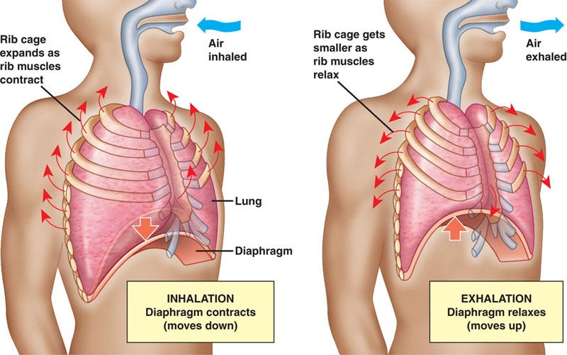
The diaphragm is a dome-shaped muscle that separates the chest cavity (thorax) from the abdomen and it’s the primary muscle in the inhalation/exhalation process. The diaphragm is the major muscle of inspiration and accounts for approximately 70% of the inhaled tidal volume in the normal individual.
During inhalation, the diaphragm contracts and flattens, creating a negative pressure within the chest cavity. This negative pressure draws air into the lungs. During exhalation, the diaphragm relaxes, and the chest cavity returns to its original size, forcing air out of the lungs.
The intercostal muscles are thin sheets of muscular fibers that run between the ribs in the costal spaces. There are two layers of intercostal muscles: internal and external. The external intercostals expand the rib cage during inspiration, while the internal intercostals are deeper and function to decrease rib cage size during expiration.
Neck muscles like the sternocleidomastoid and scalene muscles help in the inspiration process, lifting the rib cage up and allowing the lungs to expand as they let the air in.
Abdominal muscles like the rectus abdominis helps bring the rib cage down during active exhalation process.
“A highly coordinated recruitment of two or three muscle groups is required to avoid these effects. During breathing at rest, this is accomplished by the coordinated activity of the diaphragm and inspiratory rib cage muscles. Normally no expiratory muscles are used.” ~ National Library of Medicine
To understand the tiniest of structures in the body and how they work, we use microscopes and varieties of specific lenses. This is a signature called Histology that we all study in our first years of Medicine. My dad has been a Histology professor his entire professional life after graduating from Veterinary school in the 70’s. He has been teaching this stuff for ages, and his books are something else.
Since human and animal histology can be studied together to some extent, and since he’s aware I’m writing this series on the human body systems, we have been sitting down together to study and take pictures of his old histology books. These are classic books that have been discontinued. So unless you studied a medical science before the 1980’s, it’s very unlikely you'll see images like this in modern times. It’s personally valuable, and being able to bring this knowledge through my Dad into my newsletter means the world to me. I hope you love them too!
You will notice it’s all written in French language, and you’ll be able to recognise words that we have already mentioned, like bronchioles or alveoli. Most graphics done back then were simple and straightforward, sometimes requiring a big deal of imagination. Fun times! :)
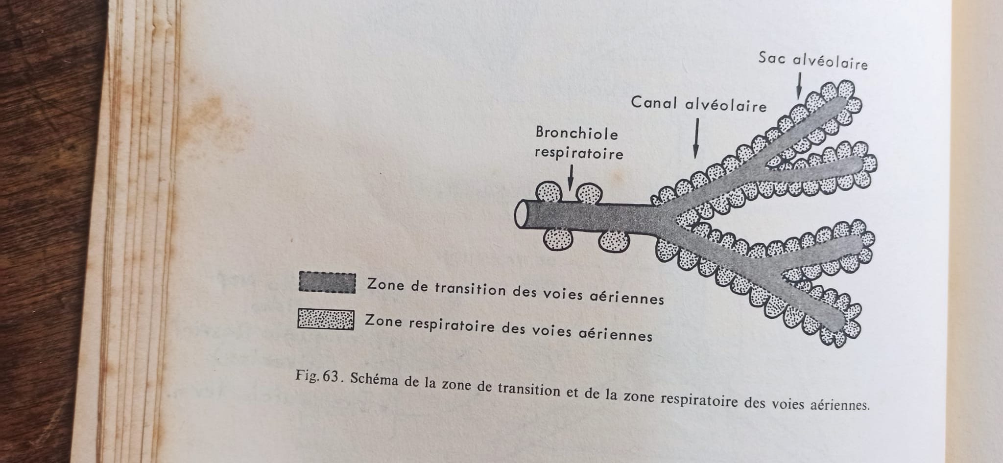
In the image above, you can see the lung’s tunnels with their names according to their location, along with the air sacks forming towards the final bits of such canals, where alveoli sacks are formed.
In the following images, you can see 1) a diagram of one lung and all its layers (pleura) which protect the lungs and help in maintaining the needed pressure for the respiration process to function. In image 2), you can see branches like a tree. That’s a diagram of the different branch sections and levels there are in the lungs. Fascinating stuff. And finally, in image 3) you can see a diagram of a lung according to the different transitions both of air and circulation, and the exact spot where lung tissue and circulatory tissue meet to allow the gas (oxygen/CO2) exchange to happen.
Bless timeless pictorial art.


How Do Heart and Lungs Connect?
Lastly, let’s see how these two main organs work together to keep us alive.
In short: the lungs oxygenate the blood that is pumped out by the heart towards your entire body for proper functioning.
Without the oxygen captured by the lungs, and without the cells carrying the oxygen through the blood (erythrocytes), there wouldn’t be human life.
The way they do this work together is fascinating, and the best way to explain it is through a video explanation. Because we’ve already studied the circulatory system (here’s a refresher if you need one before watching the following video), you’ll be able to recap some concepts and understand how lungs and heart work together.
This is the best video I have found (I’m very picky about the videos and images I share, trust me):
If you prefer a shorter version, here’s another good quality explanation on how lungs and heart work together in the body:
You can now see how circulatory and respiratory systems are deeply connected. The most notorious (and weird!) thing here, as you might have noticed, is this:
Arteries and veins are usually depicted in colours—arteries being red (oxygenated blood) and veins being blue (deoxygenated blood). That is on the normal circulation system. In the lung circulation system however, arteries are blue and veins are red.
Why? Because the blood leaving the heart into the lungs has no oxygen—meaning, it is an artery because it’s leaving the heart, and depicted in blue because it’s carrying deoxygenated blood. The same happens with the (pulmonary) vein: it’s a vein because it’s bringing blood back to the heart, however, this blood is now filled up with oxygen after travelling through the lungs, which makes it oxygenated blood, be depicted in red. Sounds tricky but if you have a read at this again, you’ll get the hang of it, I promise.

I hope this is all nice and clear!
The respiratory system is fascinating and now you know the basics: how it looks like, how it works, and how it works together with the lungs to feed you oxygen.
Next time, we will dive into what happens when the respiratory system gets sick. We will cover the most important basics on respiratory disease: ways in which it can be affected, ailments like allergies or asthma, pneumonia and more complex situations such as pulmonary fibrosis, chronic pulmonary obstruction, and lung cancer. (If there’s any particular respiratory disease you’d like me to cover, please leave a comment in the comment section below and I’ll include it as best as possible.)
As always, if you have any doubts, write it in the comments where I will reply so that everyone can learn from it too.

One Last Prompt For You
Let me give you a little respiratory prompt now that you know a great deal about your lungs:
This week, when you wake up or as you go to bed, take 30 seconds (just 30!) to stop and think about your lungs.
Slow down your breathing, take one deep inhale slowly while visualising and sensing the air that goes in. Feel as the air goes inside your nostrils—is it warm? Is it cold?
Follow the breathe inside, all the way down to your throat and into your lungs. Hold it for two seconds, and then exhale through your mouth while resounding a little “aaaah” sound out loud.
Repeat this two more times. Simply make sure to visualise the air and your lungs as they expand inside your chest.
After you’re done, say "Thank you, lungs, for this air.” See how you feel afterwards and take a mental note on it. That’s it! :)
This simple exercise might seem dull but in reality it’s complex and whole. Breathing is one of the most vital assets for human health. It impacts not just your lung capacity or your oxygen supply, but also your brain, your muscle performance, your emotions, your thought processes, and your sense of calmness.
Breathing is the most well-known grounding method since ancient times. Practices like meditation, yoga, pilates, Ayurveda and Traditional Chinese Medicine, they all have one thing in common: breathing as the main focus towards good, balanced health—a concept still clear, present, and strong today.
Have a wonderful week!
And may your air, oxygen, and peacefulness be abundant every day.
Much love,
Dr. Mariana

If you enjoyed this newsletter and want to help support it with a one-off donation of your choice, you can do here in my Tip Jar. Thank you!
Thank you for this information, Dr. M. I had no idea there were so many muscles involved with breathing! Thank you, Dad, for your input and the photos of your books. They’re easy to understand.
Dr. M., is there a lung condition for people who grew up in smoggy conditions when we were young? Air quality affects us. What can be done if we can’t avoid smog? Thank you for your work!